
Buy Pre-owned Cystoscopy Surgical Camera & Urology Surgical Camera in India — Karl Storz SPIES, Stryker Camera for TURBT, TURP & Bladder Tumor Detection
Looking for a high-performance cystoscopy surgical camera or urology surgical camera in India at an affordable price? Emcult — Noida’s most trusted pre-owned surgical equipment supplier — stocks certified pre-owned Karl Storz Image 1 SPIES Urology Camera System, Karl Storz IMAGE1 S, Stryker 1588 AIM camera, and Stryker 1688 4K AIM camera — all tested, installed, and ready for your urology operating room. Every cystoscopy surgical camera we supply is priced 40–70% below new equipment, with free installation, CSSD staff training, AMC support, and delivery across all 28 states of India.
What is a cystoscopy surgical camera?
A cystoscopy surgical camera is the specialized camera system used during cystoscopy — the procedure where a urologist inserts a thin tube (cystoscope) through the urethra into the bladder to look inside. The word “cysto” means bladder in Latin. Unlike laparoscopy (where a camera enters through the abdominal wall), cystoscopy enters the body through the natural urinary opening — making it one of the most minimally invasive diagnostic and surgical procedures in urology.
The complete cystoscopy surgical camera system consists of: a camera head that attaches to the cystoscope eyepiece and captures the image, a camera control unit (CCU) that processes the signal into HD video, a light source that sends bright illumination through the scope into the bladder, and a HD display monitor that shows the urologist the live image of the bladder wall, urethra, and ureteral orifices.
Modern cystoscopy surgical cameras from Karl Storz and Stryker go far beyond simple visualization. The most advanced systems feature NBI-equivalent technology (Narrow Band Imaging) — a technique that uses specific wavelengths of light to make flat bladder tumors and abnormal blood vessel patterns visible that would be completely missed on standard white light cystoscopy. This is clinically proven to increase bladder cancer detection rates and reduce recurrence after TURBT.
Why cystoscopy cameras are different from laparoscopy cameras
A cystoscopy surgical camera works in a very different environment from a laparoscopic surgical camera or arthroscopy surgical camera. The bladder is a fluid-filled space distended with saline irrigation — the camera must work through water rather than air. This means the light source must produce extremely bright output to penetrate the saline medium and illuminate the entire bladder dome and posterior wall. The optics must be optimized for a fluid environment with minimal glare from the water surface.
Additionally, cystoscopy surgical cameras must be compatible with both rigid cystoscopes (for TURBT, TURP, ureteroscopy) and flexible cystoscopes (for outpatient surveillance cystoscopy in office or ward settings) — two completely different scope types with different optical systems and working diameters.
The cameras used for hysteroscopy surgical camera work and those used for cystoscopy share similar requirements — both operate in fluid-filled cavities under irrigation. Emcult supplies specialized camera systems for both specialties. Similarly, laparoscopic surgical camera platforms like the Stryker 1588 AIM have a dedicated Cystoscopy specialty preset that optimizes all these parameters automatically.
The clinical importance of NBI in cystoscopy
Bladder cancer is the 10th most common cancer in India. The primary challenge in bladder cancer management is detecting flat, low-grade lesions (carcinoma in situ — CIS) and flat urothelial abnormalities that are completely invisible on standard white light cystoscopy. These lesions appear identical to normal urothelium under white light.
NBI (Narrow Band Imaging) technology — and its equivalent SPIES imaging in Karl Storz cameras — uses blue (415nm) and green (540nm) wavelengths of light that are selectively absorbed by hemoglobin. This makes the rich blood vessel network of tumors and abnormal mucosa appear dark brown/black against a normal pink-green background — making previously invisible lesions clearly visible. Multiple published studies have shown NBI cystoscopy increases detection of CIS by 20–40% compared to white light alone. For Indian urology departments dealing with high volumes of bladder cancer, this technology can directly change patient outcomes.
Urological procedures using cystoscopy surgical cameras
A urology surgical camera platform must support the full range of urological endoscopy procedures. Here is a complete guide to every major procedure that requires a cystoscopy surgical camera system.
Diagnostic Cystoscopy
Outpatient / OPD
TURBT — Transurethral Resection of Bladder Tumor
OT · Cancer surgery
TURP — Transurethral Resection of Prostate
OT · BPH surgery
Ureteroscopy (URS / RIRS)
OT · Kidney stone
PCNL — Percutaneous Nephrolithotomy
OT · Large kidney stone
Urethroscopy / Urethroplasty Assessment
Diagnostic + surgical
Laparoscopic Urology — Nephrectomy / Pyeloplasty
Laparoscopy · Urology
Bladder Biopsy / Fulguration
Diagnostic + minor OT
Karl Storz urology camera systems — available pre-owned at Emcult
Karl Storz camera systems are the global leader in urological endoscopy. Their SPIES (Storz Professional Image Enhancement System) technology — which provides NBI-equivalent imaging — was developed specifically with urology in mind, and the Karl Storz Image 1 SPIES Urology Camera System is widely regarded as the most clinically advanced urology surgical camera available for bladder tumor detection, surveillance cystoscopy, and endourology in Indian hospitals.
Karl Storz Image 1 SPIES Urology Camera System — best-in-class bladder cancer detection
The Karl Storz Image 1 SPIES Urology Camera System is specifically engineered for urological endoscopy. It combines Full HD imaging with three SPIES advanced imaging modes that dramatically improve the urologist’s ability to see what standard white light cystoscopy cannot show:
- CLARA mode: Balances the exposure across the entire bladder image — the bright central dome and the darker posterior and lateral walls are simultaneously well-exposed. This eliminates the common problem in cystoscopy where the camera overexposes the directly illuminated area while the bladder margins remain in shadow.
- CHROMA mode: Enhances color contrast between normal urothelium and abnormal tissue — making subtle mucosal changes, flat tumors, and areas of CIS appear with greater color differentiation from surrounding normal tissue.
- SPECTRA A/B mode: Narrow Band Imaging equivalent — uses specific wavelengths to highlight the abnormal vessel architecture of urothelial tumors and CIS. Flat lesions and carcinoma in situ that are invisible on white light appear as distinct dark-brown or burgundy areas against the normal background. This is the key mode for surveillance cystoscopy and TURBT mapping in bladder cancer patients.
- Advanced imaging: CLARA · CHROMA · SPECTRA
- NBI-equivalent: SPECTRA A/B mode
- Resolution: Full HD 1920×1080
- Optimized for: Cystoscopy · TURBT · TURP
- Scope compatibility: Rigid + Flexible Karl Storz
- Pre-owned price: ₹7–15 lakhs
Clinical tip: For Indian urology departments with a high bladder cancer surveillance load — the Karl Storz Image 1 SPIES Urology with SPECTRA mode can reduce the number of biopsies needed by clearly distinguishing flat CIS from normal mucosa, saving procedure time and patient discomfort.
Stryker camera systems for cystoscopy and urology — available pre-owned at Emcult
Stryker camera systems — particularly the 1588 AIM and 1688 4K AIM — provide excellent cystoscopy surgical camera performance through their dedicated Cystoscopy specialty preset, which automatically optimizes brightness, color balance, and image processing for the fluid-filled bladder environment.
Stryker 1588 AIM — advanced cystoscopy imaging with AIM modes
The Stryker 1588 AIM camera is a capable and widely used urology surgical camera platform in India. Its Cystoscopy specialty preset automatically sets optimal brightness and color for bladder visualization. The DRE (Dynamic Range Enhancement) mode is particularly useful in cystoscopy — it simultaneously brightens the posterior bladder wall and dome (which are often underexposed as the light comes from the cystoscope pointing forward) while keeping the directly illuminated anterior wall correctly exposed.
The Stryker 1588 AIM also supports IRIS mode — placing lighted ureteral stents inside the ureters to make them visible during complex laparoscopic urological procedures like laparoscopic hysterectomy, Wertheim’s radical hysterectomy, or colorectal resection where the ureters are at risk. This cross-specialty capability is extremely valuable for urology departments that also support gynecology OTs.
- Resolution: Full HD 1920×1080
- Cystoscopy preset: Dedicated optimized
- DRE mode: +150% posterior wall
- IRIS mode: Ureteral protection
- Specialties: 9 incl. Cystoscopy
- Pre-owned price: ₹8–15 lakhs
Note: The Stryker 1588 AIM does not have a dedicated NBI-equivalent mode for bladder tumor detection. For urology departments where bladder cancer surveillance and TURBT are a major part of the caseload — the Karl Storz Image 1 SPIES Urology with SPECTRA mode is the more clinically appropriate choice. For general urology (TURP, stone surgery, laparoscopic urology) — the Stryker 1588 AIM is excellent value.
Stryker 1688 4K AIM — 4K urology visualization for complex procedures
The Stryker 1688 4K AIM camera provides 4K UHD resolution that is increasingly valued in complex urological surgery — laparoscopic radical prostatectomy (LRP), laparoscopic partial nephrectomy (LPN), and retroperitoneal laparoscopic surgery where the highest possible image clarity reduces operative time and improves precision around critical vascular and neural structures. Its SPY fluorescence mode is also used in urology for renal perfusion assessment during partial nephrectomy. Available as a premium pre-owned unit from Emcult.
Rigid cystoscope
17Fr–21Fr · OT diagnosticResectoscope (TURBT / TURP)
24Fr–26Fr · OperativeFlexible cystoscope
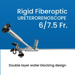
15Fr–16Fr · OPD / wardRigid ureteroscope
7.5Fr–9.5Fr · Ureter / stonesFlexible ureteroscope (RIRS)
Nephroscope (PCNL)
26Fr · Large kidney stonesCystoscopy surgical camera comparison — Karl Storz vs Stryker
Use this table to compare the leading cystoscopy surgical camera and urology surgical camera systems available pre-owned through Emcult India.
Warning: NEVER autoclave a flexible cystoscope or flexible ureteroscope — the heat destroys the internal chip-on-tip electronics and fiber bundles. Flexible scopes require High-Level Disinfection (HLD) only. Always confirm with the scope manufacturer’s IFU. Never use tap water for final rinsing — use sterile water or 70% alcohol rinse to prevent waterborne infection (Pseudomonas, Mycobacterium) in cystoscopy patients.
4mm 30° arthroscope
Standard — most commonKnee · Shoulder · Hip · Most common arthroscopy
4mm 70° arthroscope
Wide-angle for posterior accessPosterior knee · Hip (acetabulum) · Posterior shoulder
2.7mm arthroscope
Small joint arthroscopyAnkle · Wrist · Elbow · Finger joints
1.9mm arthroscope
Micro — pediatric & fingerFinger · Thumb CMC · Pediatric · Hand surgery
5mm arthroscope
Large joint — high brightnessShoulder (subacromial) · Large joint open procedures
0° arthroscope (straight)
Straight-ahead viewACL tunnels · Specific hip techniques · Teaching
How to set up a urology cystoscopy camera system — step-by-step guide for Indian hospitals
Setting up a new urology endoscopy room or OT in India requires careful selection of camera, cystoscopes, light source, and irrigation system. Here is a practical setup guide from Emcult’s biomedical engineering team.
- Choose camera based on primary urology focus: Bladder cancer–heavy caseload (TURBT, surveillance cystoscopy) → Karl Storz Image 1 SPIES Urology with SPECTRA mode. General urology (TURP, stone, flexible cystoscopy) → Karl Storz IMAGE1 S or Stryker 1588 AIM. Laparoscopic urology focus → Stryker 1688 4K AIM or Stryker 1588 AIM. No Sterrad/V-PRO → Karl Storz IMAGE1 S with H3-ZA TH104 autoclavable head.
- Select cystoscope set based on procedure mix: Full urology OT setup: 17Fr/21Fr rigid diagnostic cystoscope (0° and 30°) + 24Fr/26Fr resectoscope set for TURBT/TURP + 9Fr rigid ureteroscope + flexible ureteroscope for RIRS + nephroscope for PCNL. Outpatient urology unit: flexible cystoscope (15Fr–16Fr) + rigid diagnostic cystoscope.
- Set up irrigation system and distension media: TURP requires non-conducting isotonic glycine or sorbitol irrigation. TURBT, URS, and diagnostic cystoscopy use normal saline. Set the irrigation pressure (60–80 cmH₂O for bladder, lower for ureter). Insufficient irrigation pressure leads to a collapsed view — the camera system cannot compensate. Emcult advises on compatible arthroscopy pump / urology irrigation systems during installation.
- Configure SPIES / specialty preset on the CCU: For Karl Storz IMAGE1 S or SPIES Urology system: navigate to the SPIES menu and confirm CLARA, CHROMA, and SPECTRA modes are available. Assign SPECTRA to a quick-access camera head button for rapid mode switching during TURBT. For Stryker 1588 AIM: select the Cystoscopy specialty preset on the Home screen.
- Perform white balance before every list: Point the cystoscope at white gauze under the light source. Press and hold WB button. Wait for “WHITE BALANCE COMPLETE”. Do this at the start of every procedure list — or whenever the scope or camera head is changed. Skipping white balance causes inaccurate tissue color — abnormal mucosa may not be identifiable from normal tissue.
- TURBT protocol — NBI/SPIES first, then white light resection: Standard TURBT protocol using NBI/SPIES: (1) Insert cystoscope, fill bladder with saline. (2) Activate SPECTRA/NBI mode — systematically examine all bladder walls, dome, trigone, and lateral walls. (3) Mark or note all lesions (brown/dark on SPECTRA = tumor or abnormal vessel). (4) Switch to white light. (5) Resect all marked lesions with resectoscope. (6) Final NBI check after resection to confirm completeness. This protocol maximizes complete resection rates.
- Reprocessing — cystoscope and camera head: Karl Storz H3-ZA TH104 camera head: manual enzymatic clean → standard autoclave. Stryker 1588/1688 camera heads: manual enzymatic clean → Sterrad or V-PRO only. Rigid cystoscopes (Hopkins rod telescopes): manual enzymatic clean → autoclave. Flexible cystoscopes: manual enzymatic clean → High-Level Disinfection (HLD) in automated endoscope reprocessor (AER) using glutaraldehyde, OPA, or peracetic acid — NOT autoclave (destroys flexible scope electronics).
Warning: NEVER autoclave a flexible cystoscope or flexible ureteroscope — the heat destroys the internal chip-on-tip electronics and fiber bundles. Flexible scopes require High-Level Disinfection (HLD) only. Always confirm with the scope manufacturer’s IFU. Never use tap water for final rinsing — use sterile water or 70% alcohol rinse to prevent waterborne infection (Pseudomonas, Mycobacterium) in cystoscopy patients.
How to choose the right cystoscopy surgical camera for your Indian urology department
Factor 1 — bladder cancer caseload
If your urology department has a significant bladder cancer surveillance and TURBT workload — the Karl Storz Image 1 SPIES Urology Camera System with SPECTRA mode is the most clinically important investment you can make. SPECTRA (NBI-equivalent) imaging has been shown in multiple published studies to increase flat lesion and CIS detection rates by 20–40% compared to white light alone. For Indian urology departments where early bladder cancer detection directly impacts patient survival, this camera system provides a proven clinical advantage that justifies the investment.
Factor 2 — procedure volume and case mix
High-volume TURP and stone surgery without a major bladder cancer component → Stryker 1588 AIM camera with Cystoscopy preset is excellent value. Mix of endourology, TURBT, flexible cystoscopy, and laparoscopic urology → Karl Storz IMAGE1 S provides maximum flexibility as a single platform covering all scopes and all procedures. Complex laparoscopic radical prostatectomy, partial nephrectomy, and advanced urooncology → Stryker 1688 4K AIM for 4K visualization advantage.
Factor 3 — sterilization infrastructure
As discussed for arthroscopy cameras and laparoscopic cameras — Stryker camera heads require Sterrad or V-PRO sterilization. If your urology OT does not have a low-temperature sterilizer, the Karl Storz IMAGE1 S with H3-ZA TH104 autoclavable head is the right choice. In high-volume urology lists (10–15 cystoscopies per day), autoclavability of the camera head between cases is a significant operational advantage.
Factor 4 — cross-specialty use with hysteroscopy and laparoscopy
The cystoscopy surgical camera platform in your urology OT can often serve as the hysteroscopy surgical camera platform for your gynecology department — and the laparoscopic surgical camera for your general surgery OT. The Karl Storz IMAGE1 S and Stryker 1588 AIM both support 9 surgical specialties on one CCU — standardizing your entire hospital’s MIS OT infrastructure on a single platform and saving ₹20–40 lakhs in capital equipment.
External clinical reference
The European Association of Urology (EAU) Guidelines on Non-Muscle-Invasive Bladder Cancer recommend NBI-guided TURBT as a technique that improves detection and reduces recurrence rates — supporting the use of SPIES/NBI cystoscopy cameras in urology departments with a bladder cancer treatment program.
Why buy your urology cystoscopy camera from Emcult?
- Certified pre-owned quality: Every cystoscopy surgical camera passes our 28-point inspection — image quality, SPIES/CLARA/SPECTRA mode function, camera head buttons, cable integrity, coupler focus range, light source output, and IEC 60601-1 electrical safety testing.
- 40–70% savings: Karl Storz Image 1 SPIES Urology new costs ₹20–30 lakhs. Emcult pre-owned starts from ₹7 lakhs — same clinical SPECTRA NBI performance at a fraction of the price.
- Free installation and CSSD training: Biomedical engineers install the camera tower, configure SPIES modes, and train your urology nurses and CSSD staff on scope reprocessing — completely free with every purchase.
- TURBT protocol support: Our clinical specialist team advises your urology consultants on the NBI/SPIES TURBT protocol — maximizing the clinical benefit of the camera’s advanced imaging modes from day one.
- AMC and 24/7 support: Annual Maintenance Contracts covering preventive maintenance, camera head leak-down testing, light source output verification, and 24/7 emergency breakdown support across all 28 states.
- GEM/DGSND procurement: Government urology departments and medical college urology units can procure through GEM portal with full DGSND rate contract support, CE certificates, and GST invoices.
Frequently asked questions — cystoscopy & urology surgical camera India
What is the price of a cystoscopy surgical camera in India?
What is SPIES imaging in Karl Storz cystoscopy cameras?
Which cystoscopy camera is best for TURBT in India?
Can I use the same camera for cystoscopy and laparoscopic urology?
Can a flexible cystoscope be sterilized in an autoclave?
What is the difference between NBI and SPIES in urology cameras?
Does Emcult provide AMC for urology cystoscopy camera systems across India?
₹3.5 – 35 Lakhs
- Laparoscopy Surgical camera
- Arthroscopy Surgical Camera
- ENT Surgical Camera
- Hysteroscopes Surgical Camera
- Urology Surgical camera
- Flexible endoscopes
- Colonoscope
- Bronchoscope
- ERCP duodenoscope
+91 971-788-7658
emcult7@gmail.com
We Delivered Laparoscopic Surgical Camera in Pan India:
- Cystoscopy Surgical Cameras in Delhi NCR
- Cystoscopy Surgical Cameras in Pryagraj
- Cystoscopy Surgical Cameras in Mumbai
- Cystoscopy Surgical Cameras in Bengaluru
- Cystoscopy Surgical Cameras in Chennai
- Cystoscopy Surgical Cameras in Hyderabad
- Cystoscopy Surgical Cameras in Kolkata
- Cystoscopy Surgical Cameras in Lucknow
- Cystoscopy Surgical Cameras in Jaipur
- Cystoscopy Surgical Cameras in Ahmedabad
- Cystoscopy Surgical Cameras in Pune
- Cystoscopy Surgical Cameras in Chandigarh
- Cystoscopy Surgical Cameras in Bhopal
- Cystoscopy Surgical Cameras in Guwahati
- Cystoscopy Surgical Cameras in Kochi
- Cystoscopy Surgical Cameras in Bhubaneswar